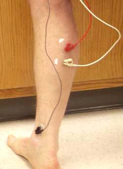
| In this lab, you record an electromyogram (EMG) of your gastrocnemius muscle with surface electrodes. The positive and negative recording electrodes (red and white) are placed on the back of the calf and the ground electrode (black) is attached to the ankle. The spiky blue trace in the picture above shows the electrical activity of the muscle during walking. |
 |
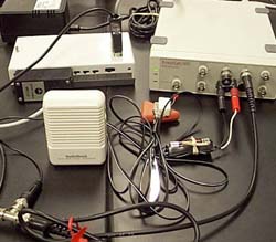
| The electrode leads are attached to the differential amplifier ( upper left). The amplified signal then feeds into the PowerLab data acquisition system (upper right) and audio monitor (left), which allows you to hear the EMG as well as see it. |
 |
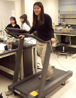
| You can record an EMG while standing, sitting, jumping, or walking on the treadmill to investigate the role of the gastrocnemius muscle in these activities. |
 |
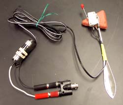
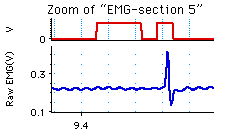
| With this percussion hammer, you can tap the achilles tendon to stimulate a reflex. The inertial switch on the head of the hammer causes a 1.5 V electric pulse when the hammer strikes. This can be recorded on the computer to mark the time of the tendon tap. |
 |
| In this trace the beginning of the first red pulse represents the tendon tap and the blue spike is the EMG response. The delay between these two represents the conduction time. You can use this to calculate the conduction velocity in the reflex arc if you know the distance the signal travels along the nerves. |
 |