Home
Lab Tour
Lab Syllabus
Papers
Symposium
 Resources Resources
-
-
|
-

Lab 8: Human Electrocardiography
[Reading] [Overview] [Useful Links] [Lab Report] [Further Study]
Pre-lab reading
- Lab manual - pages 75-90
- Vander, Sherman, and Luciano - Chapter 14, especially sections C and F.
Overview
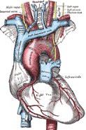
The vertebrate heart is a muscular pump capable of functioning without influence from the nervous system. As you may have discovered last week, the frog heart can beat rhythmically and pump fluid for awhile even after removal from the body, as long as it is well oxygenated. This independence from direct nervous control relies on two critical features of cardiac muscle: (1) the ability to generate a rhythmic pattern of contractions, and (2) control of the timing of contraction of different parts of the heart so that blood is moved in a coordinated fashion from one chamber to another in the right order. These features involve generating and conducting action potentials in specialized muscle cells, resulting in electrical currents following a complex path through the heart. The electrocardiogram (ECG, or EKG after the German electrokardiogram) is a recording of these electrical signals from electrodes placed (usually) on the skin surface. A typical ECG uses multiple electrodes placed on the chest and limbs to "view" the electrical signals from up to 12 different perspectives. We will focus on one of the most commonly used ECG leads to examine the major events in the cardiac cycle.
This lab exercise consists of three sections focusing on different questions:
- Record a resting ECG. How do electrical signals on the trace correlate with events in the heart? What are the relative proportions of systole and diastole in the heartbeat? How regular is the heart rhythm?
- Record an ECG on the same person during exercise. As heart rate increases, which components of the cardiac cycle are shortened? Does the relative proportion of systole and diastole change? Does it matter?
- How does the ECG reveal abnormalities in heart function? Since we are unlikely to observe major abnormalities in our small (and, we hope, healthy) sample of the population, we'll provide case histories of people with unusual ECGs and ask you to analyze the differences and speculate on the possible causes.
Useful Links
- EKG Tutorial from NYU Medical School. Very clear explanation of electrical basis for ECG and outstanding animations comparing electrical recording to the spread of excitation in the heart.
- Visit the McGill University Virtual Physiology Lab and click on their Cardiovascular lab for an excellent tutorial on measuring and understanding electrocardiograms.
- 12-Lead ECG Library - A collection of ECG images with explanations of the associated heart conditions
- ECG Rhythms - a dictionary of heart abnormalities (fairly technical).
Lab Report
No lab report due for this lab.
Further Study at Berkeley
Courses related to this lab topic
IB129. Human Physiological Assessment. Principles and theories of human physiological assessment in relation to physical activity and conditioning. Performance of laboratory procedures in the measurement and interpretation of physiological fitness (cardiorespiratory endurance, body composition, musculoskeletal fitness).
(Johannessen)
IB129L. Stress Testing Physical Assessment. Clinical application and practice of exercise testing, electrocardiography, and exercise prescription in UC Fitness Evaluation Program.
Faculty doing research related to this lab:
- George A. Brooks (IB) Exercise physiology and metabolism.
- Sue Johannessen (Phys.Ed.) Fitness training and assessment.
-
Links on these pages to commercial sites do not represent endorsement by the University of California or its affiliates.
IB 132 Lecture Website
U. C. Berkeley
Send us your comments on the IB132L pages!
-
-
Last updated 1/13/06
- Copyright © Department of Integrative Biology. All rights reserved 2006
|
|